Oxidative stress & single-cell biology
Understanding heterogeneous bacterial responses at the level of individual cells.

Research
Our laboratory investigates how light-generated reactive oxygen species interact with microorganisms, biological interfaces, and engineered materials. We combine mechanistic photochemistry with quantitative microbiology and imaging to understand oxidative stress and develop new antimicrobial strategies.
Research directions
Each research direction addresses a different scale of the same central problem: how reactive oxygen species can be generated, controlled, measured, and used to influence microbial systems.
Understanding heterogeneous bacterial responses at the level of individual cells.
Connecting photosensitizer photochemistry with microbial inactivation mechanisms.
Combining biological targeting with photochemical generation of oxidative stress.
Developing reusable light-responsive platforms, including 3D-printed systems.
Research program
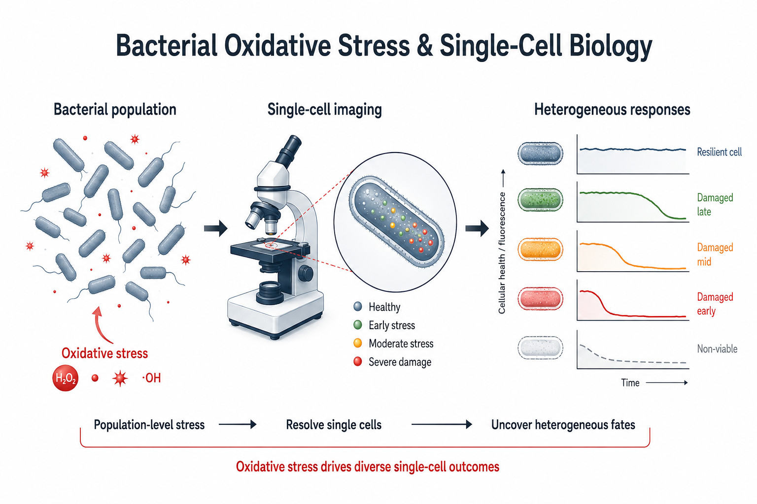
Genetically similar bacteria can respond very differently to the same oxidative challenge. Population-level measurements often hide this variability, making it difficult to understand why some cells are rapidly damaged while others persist.
We use fluorescence microscopy and quantitative image analysis to follow individual cells during photodynamic treatment. These experiments allow us to measure the timing and distribution of membrane permeabilization, oxidative damage, and survival-related phenotypes across bacterial populations.
Time-resolved imaging of bacterial viability, membrane integrity, and stress responses.
Extraction of cell-level kinetic parameters from microscopy datasets.
Analysis of how physiological state changes the distribution of oxidative-stress responses.
Connecting bulk antimicrobial outcomes with cell-to-cell heterogeneity.
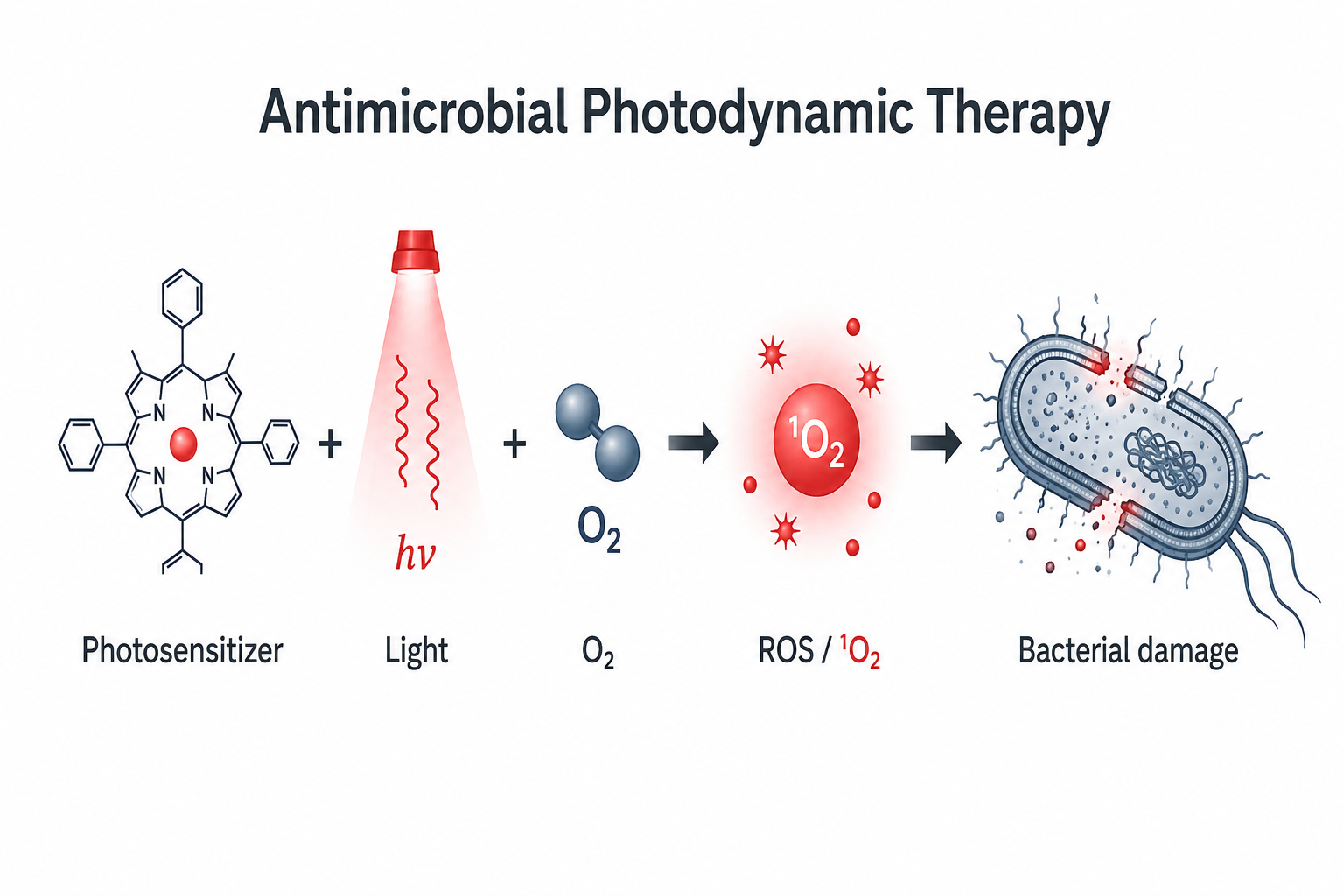
Antimicrobial photodynamic therapy uses a photosensitizer, light, and molecular oxygen to generate reactive oxygen species capable of damaging microbial cells. Our work focuses on understanding the photochemical and biological factors that determine treatment efficacy.
We evaluate photosensitizer performance using complementary spectroscopic, chemical, microbiological, and imaging approaches. A major goal is to distinguish improved light harvesting from changes in intrinsic reactive-oxygen generation and to connect these photophysical properties with biological activity.
Absorption, emission, photostability, and light-harvesting measurements.
Optical and chemical probes for evaluating photosensitized oxidation.
Quantitative measurements of bacterial inactivation as a function of light dose and treatment conditions.
Real-time visualization of cell damage during light exposure.
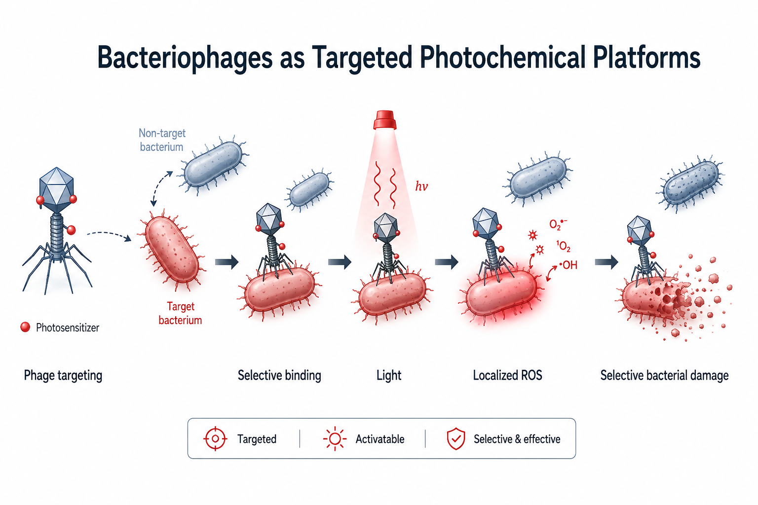
Bacteriophages provide highly specific recognition of bacterial hosts. We are exploring ways to combine that biological specificity with photosensitizer chemistry so that light-generated oxidative stress can be delivered more selectively.
This work includes studies of photosensitizer–phage association and conjugation, preservation of phage infectivity after chemical modification, and the effect of oxidative stress on phage–host interactions.
Strategies for attaching photosensitizers to phage capsids while minimizing disruption of infective structures.
Buffer exchange, centrifugal filtration, and characterization of modified phage preparations.
Monitoring bacterial growth, lysis, and infection dynamics under oxidative conditions.
Using optical reporters to probe membrane and oxidative changes during infection.
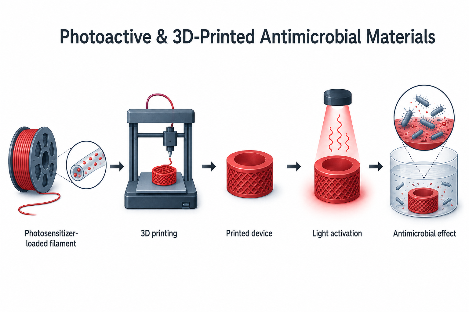
We incorporate photosensitizers into polymeric materials to create reusable surfaces and devices that respond to visible light. Additive manufacturing allows us to control geometry, composition, and application-specific design while retaining photochemical function.
Current work includes dye-loaded PLA systems produced by melt processing and 3D printing. We evaluate their optical properties, photosensitizer release, photostability, recyclability, and light-dependent antimicrobial performance.
Photosensitizer incorporation by melt blending, filament extrusion, and 3D printing.
Absorption and fluorescence measurements of photosensitizer-loaded materials.
Quantitative analysis of photosensitizer release from printed materials into aqueous media.
Light-dependent inactivation assays using printed photoactive devices and surfaces.
Cross-cutting capabilities
The lab is built around combinations of chemistry, quantitative imaging, microbiology, and materials characterization rather than a single experimental platform.
Light-driven reactions, photosensitizers, and reactive oxygen species generation.
Absorption, fluorescence, and quantitative optical characterization.
Live-cell and time-resolved imaging at the single-cell level.
Culture-based viability, growth kinetics, and antimicrobial response measurements.
Filament preparation and 3D printing of functional photoactive materials.
Kinetic modeling, image quantification, and statistical analysis across biological scales.
Work with us
Our projects provide opportunities for undergraduate and graduate students to work across chemistry, microbiology, microscopy, quantitative analysis, and materials science.