Research Projects
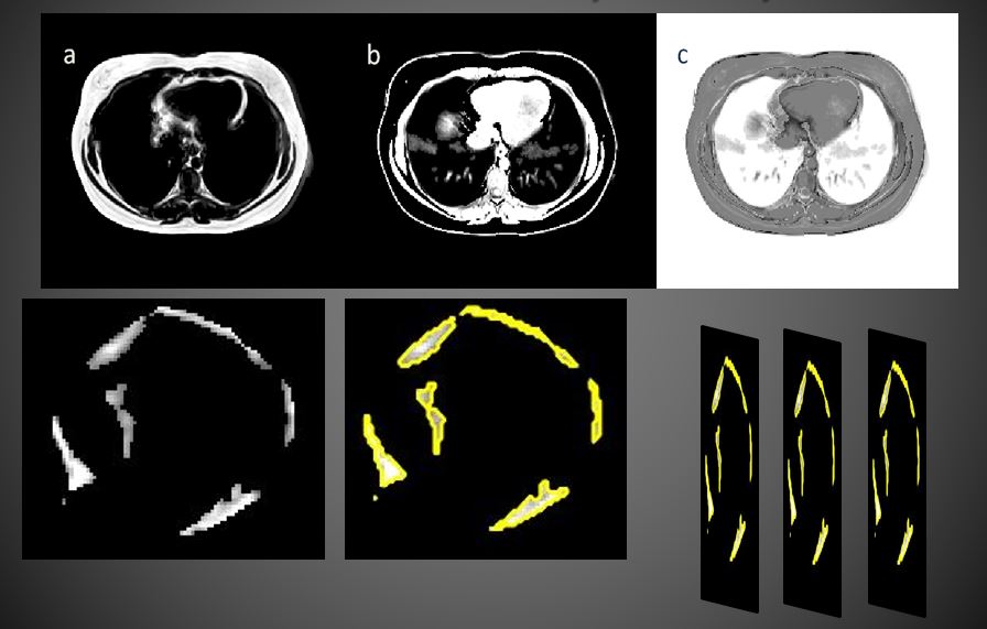
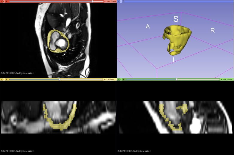
1) 3D Segmentation of Epicardial Adipose Tissue
from Cardiac MRI
Obesity is a wide-ranging health problem and fat
depots around the heart have been linked to the risk
of cardiovascular disease. Magnetic Resonance
Imaging (MRI) has emerged as the standard for
quantifying epicardial fat for clinical studies but
requires the analysis of a large number of images in a
short time. We work on 3D segmentation of
epicardial adipose tissue (EAT) and the development of
tools to aid in studies of EAT and its relationship to
cardiovascular disease and risk.
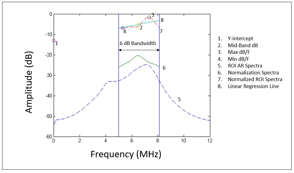
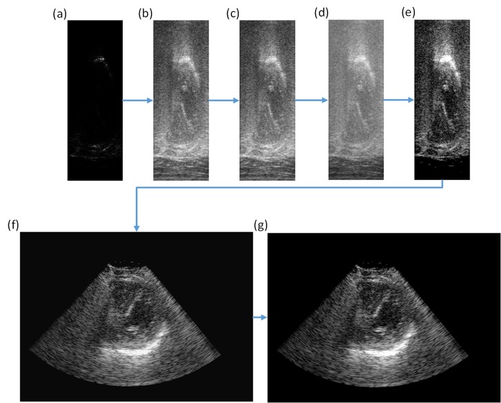
2) Identification of Epicardial Adipose Tissue
Using Echocardiography
Magnetic resonance imaging (MRI) can provide
three-dimensional (3D) assessment of EAT, but it is
expensive, time-consuming, and is only available at
large institutions.
Echocardiography is safe, real-time, inexpensive, and
can also be used to quantify cardiac structure and
function. The goal of this project is to utilize 3D
volumetric information from MRI data to develop a
shape-based model to be used in conjunction with
real-time echocardiography and advanced processing of
the radio-frequency (RF) ultrasound signals for
volumetric assessment of EAT. Machine learning
algorithms are used to differentiate tissue types based
on features from the ultrasound spectra. Leveraging the
specific individual strengths of MRI and
echocardiography has the potential to yield a more
powerful, yet less expensive analysis tool suited for
large studies of intervention and their effect on EAT
and cardiovascular health.
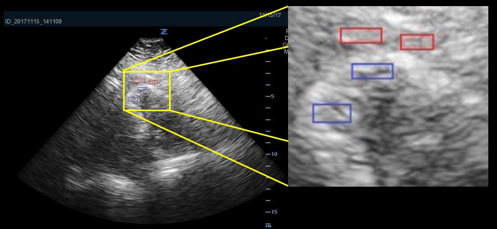
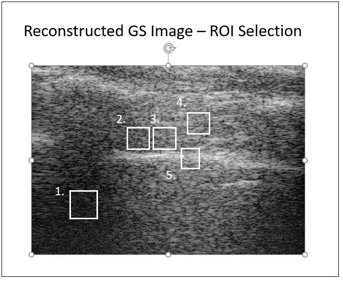
3) Spectral Analysis of Ultrasound
Radiofrequency Signals for the Identification of
Intercostal Nerves
Ultrasound has long been used to assess and image
soft-tissue structures
in vivo.
However, it
has also been used to assess mechanical properties of
tissue via elasticity imaging techniques and algorithms.
Acoustic
Radiation Force Impulse (ARFI) imaging can be used to
both mechanically excite the tissue and observe the
response of the tissue.
Images can be created to highlight the mechanical
properties of different tissues, thus providing contrast
between structures difficult to discern via traditional
b-mode imaging.
More
specifically, the project is investigating the
feasibility of imaging intercostal nerves during
image-guided procedures performed by anesthesiologists.
It is a
collaboration with the Departments of Biomedical
Engineering and General Anesthesiology at the Cleveland
Clinic Foundation.